Pulmonary Venous Congestion
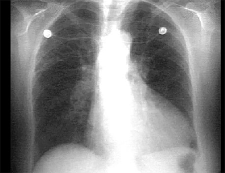
This PA chest X ray shows pulmonary venous congestion. The congestion is demonstrated by enlarged hilar shadows with increased pulmonary venous markings, especially in the upper lobes, that is, cephalization. The dilated smaller vessels can be followed all the way to the periphery. The heart size is large, with a cardiothoracic ratio greater than fifty percent. There is a prominent left ventricle. Note the monitoring electrodes can be seen on the X ray.