You are incorrect - the best interpretation of the chest X ray in our patient is pulmonary edema with a normal heart size.
Your choice: Pulmonary infiltrate
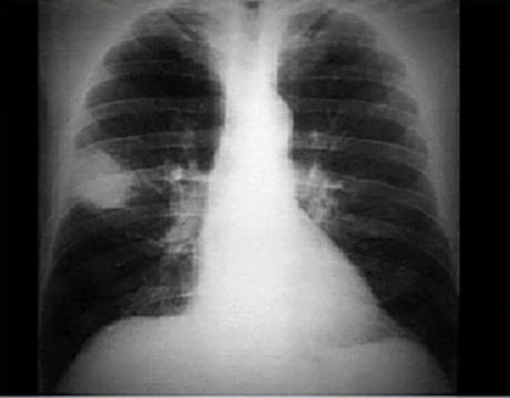
PA
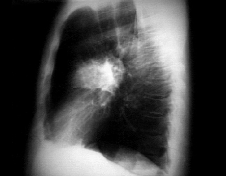
Lateral
These chest X rays show a pulmonary infiltrate.
In this PA view, there is a peripheral 6 x 4.5 cm radiodensity or infiltrate in right upper lobe. The infiltrate is free of any contained calcification or stranding into the adjacent pulmonary parenchyma. Atelectasis and signs of hilar adenopathy are also absent. The cardiac silhouette, pulmonary vasculature and bony structures are normal.
In this lateral view, the radiodensity or infiltrate is superimposed on the hilar area and can be localized to the anterior segment of the right upper lobe.