Pericardial calcification PA and Lat
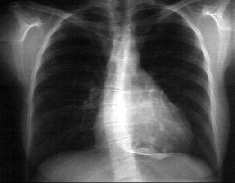
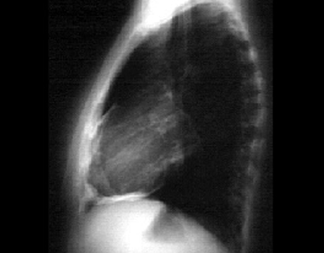
These chest X rays show pericardial calcification.
This PA view demonstrate the dense white pericardial calcification particularly well seen on the diaphragmatic surface of the heart. Note that the cardiothoracic ratio
is 50%, that is, upper limits of normal. Pericardial calcification occurs in the setting of longstanding inflammatory disease.
Note also the scoliosis of the spine.
This lateral view demonstrates the dense pericardial calcification. It is well seen on the anterior and the diaphragmatic surfaces of the heart.