You are incorrect - the best interpretation of the rhythm strip in our patient is sinus rhythm with premature atrial and ventricular complexes.
Your choice: Interpolated premature ventricular complexes
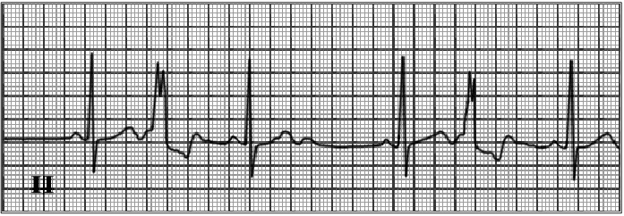
This rhythm strip shows sinus bradycardia with interpolated premature ventricular complexes (PVCs). The underlying sinus bradycardia is reflected by normal P waves preceding each sinus QRS at a rate of forthy beats per minute. Note the prominent, but normal U wave following the T wave in the normal sinus beat. These are frequently present when the rate is slow. The premature QRS complexes are wide and different from the sinus QRS and are not preceded by P waves. They are interpolated, that is, they are located between two normal sinus complexes. When the sinus rate is faster, the P wave following the ectopic beat often finds the ventricle refractory. This results in a full compensatory pause.