Cardiomegaly, pulmonary venous congestion and pleural effusion PA and Lateral
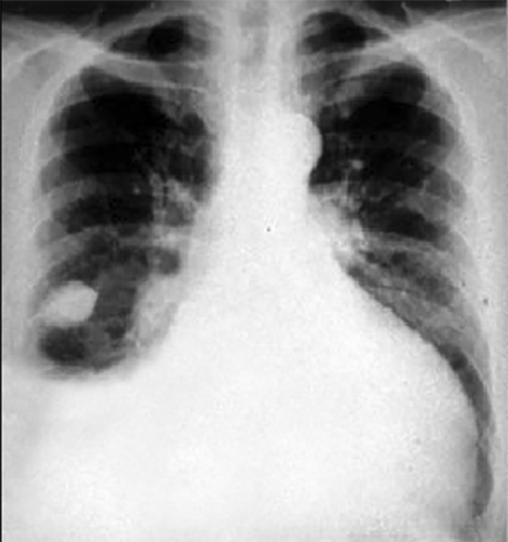
These chest X rays show cardiomegaly and pulmonary venous congestion. In this PA view, cardiomegaly is evidenced by a cardiothoracic ratio greater than fifty percent. The enlarged left ventricle is seen as an increase in the inferolateral cardiac border. Pulmonary venous congestion is demonstrated by the enlarged hilar shadows with increased pulmonary markings. These dilated smaller vessels can be followed all the way to the periphery. A large pleural effusion is demonstrated in the lower right lung field. There is loculated fluis in the right interlobar fissure. This is called a phantom tumor, as it may disappear with therapy. Its borders are perfectly smooth, oval in shape with little beaks at eigher end as the fluid trails off. These characteristics distinguish it from a true tumor.
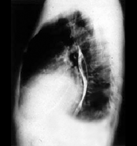
The lateral view with barium swallow shows posterior displacement of the barium filled esophagus by the
enlarged left atrium.