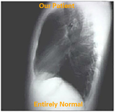
Entirely Normal
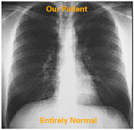
Let us analyze the borders of the cardiac silhouette beginning with the PA view.
The terms right and left always refer to the patient's right and left side. The right border consists of the aortic arch, the superior vena cava and the right atrium inferiorly. The left border consists of the ascending aorta, the main pulmonary artery, the left atrial appendage that is best seen in pathologic circumstances, and the left ventricle that comprises the most inferior portion. In the lateral chest X ray, anterior aspect of the cardiac silhouette is made up of: the ascending aorta, the right ventricular outflow tract and the right ventricle inferiorly. The posterior margin of the cardiac silhouette is formed by the left atrium above and the left ventricle below.
The terms right and left always refer to the patient's right and left side. The right border consists of the aortic arch, the superior vena cava and the right atrium inferiorly. The left border consists of the ascending aorta, the main pulmonary artery, the left atrial appendage that is best seen in pathologic circumstances, and the left ventricle that comprises the most inferior portion. In the lateral chest X ray, anterior aspect of the cardiac silhouette is made up of: the ascending aorta, the right ventricular outflow tract and the right ventricle inferiorly. The posterior margin of the cardiac silhouette is formed by the left atrium above and the left ventricle below.