You are incorrect - the best interpretation of the chest X rays in our patient is right ventricular enlargement, prominent pulmonary arteries and decreased peripheral markings.
Your choice: RV enlargement, small pulmonary trunk and ↓ pulmonary vascularity
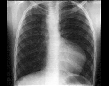
These chest X ray shows right ventricular enlargement, a small pulmonary trunk and decreased pulmonary vascularity. The PA view suggests right ventricular enlargement, as evidenced by the upturned apex. The small pulmonary trunk is evidenced by the absent convex shadow in the left hilar area. Pulmonary vascularity is also diminished, as evidenced by the absence of distal vascular lung markings. These findings are characteristic of tetralogy of Fallot. Note also the right-sided aortic arch demonstrated by the vascular density along the upper right heart border and the displacement of the trachea to the left. Right-sided aortic arch is seen in about twenty-five percent of patients with tetralogy of Fallot.