You are incorrect - the chest X rays in this patient demonstrate left atrial enlargement, biventricular enlargement and increased pulmonary arterial vascularity.
Your choice: RV Enlargement, Dilated Pulmonary Trunk, ↑ Pulmonary Arterial Vascularity - PA and Lat
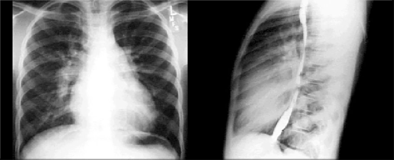
These chest X rays show right ventricular enlargement, a dilated pulmonary trunk and increased pulmonary arterial vascularity. Right ventricular enlargement is suggested in the PA view by the upturned apex and the minimally increased cardiothoracic ratio. It is suggested in the lateral view by obliteration of the retrosternal air space. The PA view shows dilation of the pulmonary trunk, manifested by the convex density below the aortic knob. Both views show prominence of the pulmonary artery, particularly well seen in the PA view as dilation of the right pulmonary artery. Increased pulmonary arterial vascularity is reflected further by prominent distal arterial markings. These chest X rays are consistent with a large atrial level left-to-right shunt, as in an atrial septal defect.