You are incorrect - this patient's electrocardiogram is within normal limits.
Your choice: Left ventricular hypertrophy
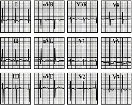
This electrocardiogram taken from a thirteen-year-old with aortic valve disease, indicates left ventricular hypertrophy. Note the precordial leads are recorded at ½ standard.
The features illustrating left ventricular hypertrophy for this age include deviation of the QRS axis into the left superior quadrant, or left axis deviation, as reflected by the dominant R wave in lead I and the dominant S wave in lead aVF and an ↑ 60 mV R wave in precordial lead V6, along with wirh repolarization changes, negative ST segments and biphasic T waves in the left ventricular leads I, V6 and V7.
 Note the precordial leads are recorded at ½ standard.