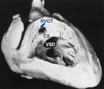
Tetralogy of Fallot Pathology Specimen
This photograph of a especially prepared fixed specimen illustrates a typical example of tetralogy of Fallot. Note the large ventricular septal defect (VSD) and the short anterior displaced conus septum (CS) severely narrowing the right ventricular outflow tract (RVOT). Note also the thickness of the right ventricular wall.