You are incorrect - the best interpretation of the chest X ray in our patient is RV enlargement + small pulmonary trunk + ↓ pulmonary arterial vascularity + right Ao arch.
Your choice: RV enlargement + small pulmonary trunk + ↑ pulmonary arterial vascularity + left Ao arch
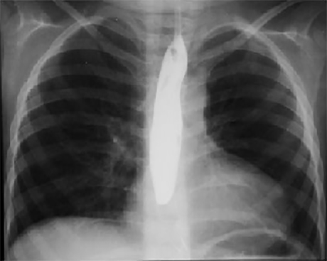
This chest X ray taken during a barium swallow shows right ventricular enlargement, a small pulmonary trunk, increased pulmonary arterial vascularity and a left aortic arch.
Right ventricular enlargement is demonstrated by the upturned cardiac apex. Decreased size of the pulmonary trunk is suggested by the straight left cardiac silhouette. Increased pulmonary arterial vascularity is reflected by
dilated central pulmonary arteries. A left aortic arch is indicated by deviation of the tracheal air shadow to the right and presence of the aortic knob at the left upper mediastinal border.