You are incorrect - the best interpretation of the chest X ray in our patient is RV enlargement + small pulmonary trunk + ↓ pulmonary arterial vascularity + right Ao arch.
Your choice: BVE + large pulmonary trunk + ↑ pulmonary arterial vascularity + left Ao arch PA and Lateral
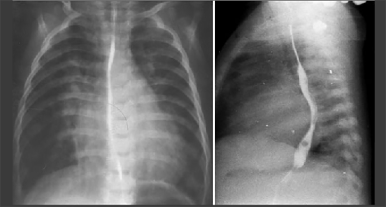
These chest X rays taken during a barium swallow show biventricular enlargement, a large pulmonary trunk, increased pulmonary arterial vascularity and a left aortic arch.
Biventricular enlargement is reflected by the large cardiac shadow extending both leftward and rightward.
The PA view demonstrates dilation of the pulmonary trunk manifested by the convex density below the aortic knob.
Increased pulmonary arterial vascularity is reflected by dilated central pulmonary arteries and prominent distal arterial vascular markings.
A left aortic arch is indicated by deviation of the tracheal air shadow to the right and presence of the aortic knob at the left upper mediastinal border.
Note also left atrial enlargement indicated in the lateral view by indentation and posterior deviation of the barium filled esophagus.