You are incorrect - the best interpretation of the chest X ray in our patient is RV enlargement + small pulmonary trunk + ↓ pulmonary arterial vascularity + right Ao arch.
Your choice: RV enlargement + large pulmonary trunk + pulmonary venous congestion + left Ao arch
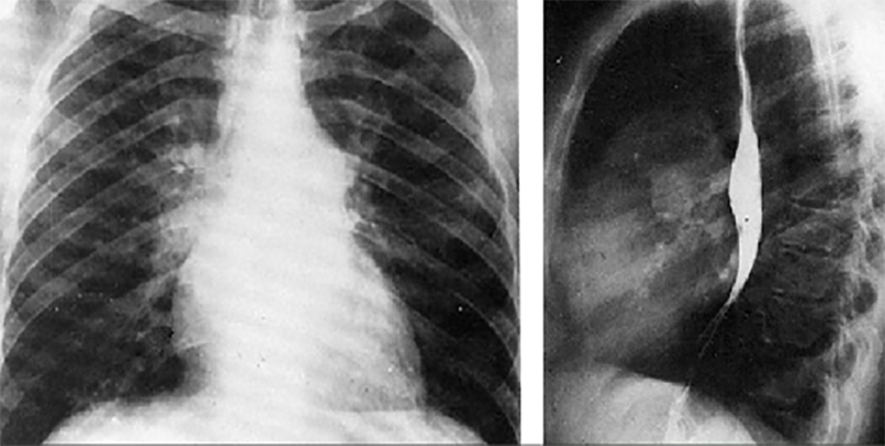
These chest X rays taken during a barium swallow show right ventricular enlargement, a large pulmonary trunk, pulmonary venous congestion and a left aortic arch.
In this PA view, right ventricular enlargement is demonstrated by the upturned cardiac apex.
The PA view demonstrates dilation of the pulmonary trunk manifested by the convex density below the aortic knob.
Pulmonary venous congestion is suggested by prominent, fine vascular markings seen in the upper lung fields.
The left aortic arch is indicated by deviation of the tracheal air shadow to the right and presence of the aortic knob at the upper left mediastinal border.
In the lateral view, right ventricular enlargement is demonstrated by loss of the retrosternal air space.