You are incorrect - this patient's electrocardiogram demonstrates biventricular hypertrophy and left atrial enlargement.
Your choice: Biventricular hypertrophy with left anterior hemiblock pattern
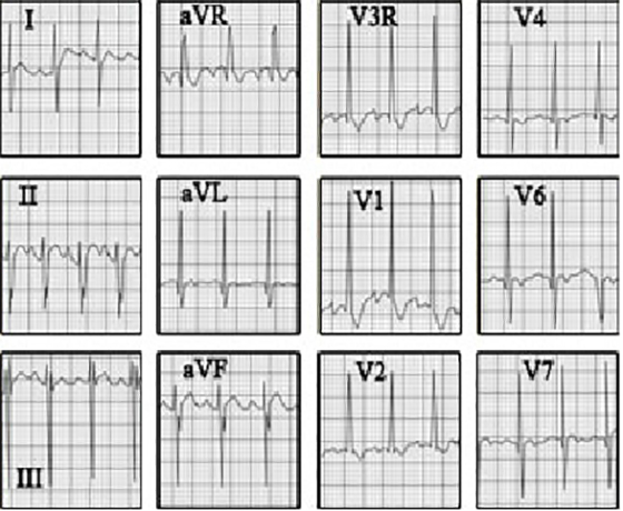
This electrocardiogram shows biventricular hypertrophy with left anterior hemiblock pattern.
The characteristic features of right ventricular hypertrophy demonstrated here include tall, dominant R waves in precordial leads V3R, V1 and V2.
The characteristic features of left ventricular hypertrophy in the presence of right ventricular hypertrophy include signigicant Q waves and dominant R waves in precordial leads V6 and V7.
The characteristic features of left anterior hemiblock pattern include a markedly superior QRS axis, as demonstrated by predominantly negative QRS complexes in leads II, III and aVF.
And initial Q waves in leads I and aVL, with the Q in aVL being larger.
The QRS axis is directed slightly to the right, as commonly occurs when the ventricles are equally affected. This type of electrocardiogram is characteristic of a complete atrioventricular canal defect, one type of endocardial cushion defect. Note that this ECG also demonstrates biatrial enlargement.