You are incorrect - this patient's electrocardiogram demonstrates biventricular hypertrophy and left atrial enlargement.
Your choice: Right ventricular hypertrophy and right atrial enlargement
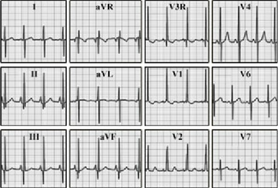
This electrocardiogram in a one-year-old infant demonstrates right ventricular hypertrophy and right atrial enlargement.
The characteristic features of right ventricular hypertrophy include right axis deviation of the QRS, reflected by predominantly negative complexes in lead I.
QR pattern with positive T waves
in precordial lead V3R.
And tall 100% R waves with positive T waves in lead V1.
The characteristic feature of right atrial enlargement is the 4 mm, peaked P waves in leads II and aVF.
