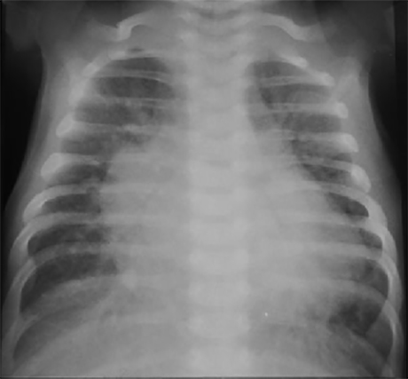
Infant with Large VSD
This is the PA chest X ray of a 3-month-old infant with a large ventricular septal defect. It demonstrates increased pulmonary vascular markings and generalized cardiomegaly.
Characteristic features include prominent vascular densities extending out from the mid line reflecting large central pulmonary arteries.
Large cardiac silhouette
reflecting enlargement of both right and left heart chambers.
And prominent upper left heart border reflecting a dilated pulmonary trunk.
Note also pulmonary hyperinflation,
reflected by both diaphragms seen at the level of the eleventh rib.
