You are incorrect - this patient's electrocardiogram is within normal limits.
Your choice: Biventricular hypertrophy
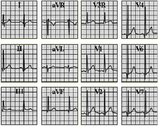
This electrocardiogram from a three-year-old patient with a large ventricular septal defect and moderate pulmonary valve stenosis shows biventricular hypertrophy.
The characteristic feature of right ventricular hypertrophy demonstrated here is dominant R waves, R/S ratio greater than one, and positive T waves in leads V3R and V1. With right ventricular hypertrophy alone the tracing should also demonstrate right axis deviation and dominant S waves, R/S ratio less than one, in leads V4 through V7. These findings are absent, because of concomittant left ventricular hypertrophy.
The characteristic features of left ventricular hypertrophy demonstrated here include prominent Q waves and tall R waves in leads V4 through V7. Note also that the QRS axis is normal at plus sixty degrees, as identified by the isoelectric lead aVL and the dominant R waves in inferior leads II, III and aVF. Note also that the six standard limb leads are normal.